Cryosonic | Professional CryoPen Lesion Removal Specialists UK
Why Early Assessment Matters for Sun-Damaged Skin Patches
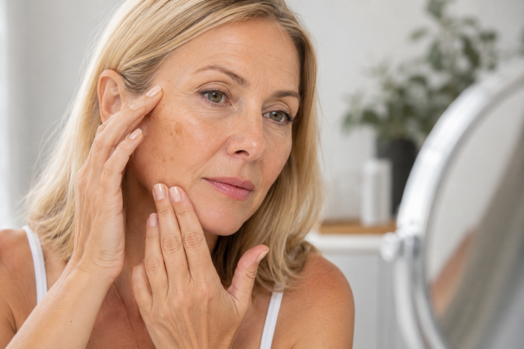
A rough patch that refuses to disappear is often dismissed as dry skin or the result of aging. Yet persistent changes on sun-exposed areas deserve closer attention, particularly when they become scaly, crusted, or sensitive. Years of ultraviolet (UV) exposure gradually age skin cells, and those changes can sometimes develop into precancerous lesions that require professional assessment.
Regular skin examinations help distinguish harmless pigmentation from conditions that may benefit from clinical management. Detecting abnormalities early often leads to simpler treatment options and better cosmetic outcomes.
Which Skin Changes Should Never Be Ignored?
Actinic keratoses commonly develop on parts of the body that receive the greatest amount of sunlight, including the face, ears, scalp, forearms, neck, and the backs of the hands. While these lesions vary in appearance, several warning signs are frequently seen:
- Rough, sandpaper-like texture
- Persistent red, pink, brown, or skin-colored patches
- Dry or flaky areas that repeatedly return
- Mild tenderness, itching, or burning
- Thickened or crusted skin that does not heal normally
Although not every lesion progresses, some may develop into squamous cell carcinoma , making professional evaluation an essential step rather than a precaution.
How Are Precancerous Sun Lessons Evaluated?
Assessment usually begins with a detailed review of medical history, cumulative sun exposure, and changes noticed over time. Clinicians examine the lesion's color, borders, thickness, and surrounding skin before deciding whether further investigation is needed.
If the appearance raises concern, a skin biopsy may be recommended to confirm the diagnosis and exclude invasive skin cancer. In many straightforward cases, diagnosis can be made through clinical examination alone.
For isolated lesions, actinic keratosis treatment may involve carefully controlled cryotherapy that freezes abnormal cells while preserving nearby healthy tissue. Other management options include topical medications, photodynamic therapy, or minor surgical procedures depending on the number, location, and characteristics of the lesions.
Treatment selection is rarely based on appearance alone. Factors such as lesion size, recurrence, immune status, previous skin cancers, and overall skin health all influence the most appropriate clinical approach.
Protecting skin after treatment
Removing visible lesions does not eliminate the underlying effects of long-term UV exposure. New areas of damage may develop over time, making prevention an ongoing part of skin care.
Daily habits that support healthier skin include:
- Apply broad-spectrum SPF 30 or higher sunscreen throughout the year
- Wearing wide-brimmed hats and protective clothing during prolonged outdoor activity
- Avoiding peak UV hours where practical
- Performing routine self-examinations to identify new or changing lesions
- Scheduling periodic professional skin checks if there is a history of significant sun exposure
People who spend years working outdoors or who have experienced frequent sunburns often benefit from continued monitoring even after successful treatment.
Why Modern Cryotherapy Remains a Common Choice
Advances in cryotherapy have made targeted treatment more precise than earlier freezing methods. Devices that deliver controlled freezing temperatures can minimize damage to surrounding tissue while effectively destroying abnormal cells.
This approach is frequently chosen for small, well-defined lesions because treatment sessions are brief and recovery is generally straightforward. Mild redness, blistering, or temporary scabbing may occur as the skin heals, with fresh tissue gradually replacing the treated area.
Clinical decisions should always balance effectiveness, cosmetic outcome, patient comfort, and the likelihood of recurrence. A personalized assessment allows healthcare professionals to determine whether cryotherapy, topical therapies, light-based treatments, or another intervention offers the most suitable option for each individual.
A Practical Perspective
Long-term sun exposure leaves lasting effects that often appear gradually rather than all at once. Paying attention to persistent rough patches and seeking timely assessment can reduce the likelihood of more serious skin changes developing unnoticed. Combining professional evaluation with consistent UV protection remains one of the most effective strategies for maintaining healthy skin throughout later life.
Verantwortlich für den Inhalt dieser Seite ist ausschließlich der
Autor dieser Homepage, kontaktierbar über dieses Formular!